Renal pathology
Hydronephrosis
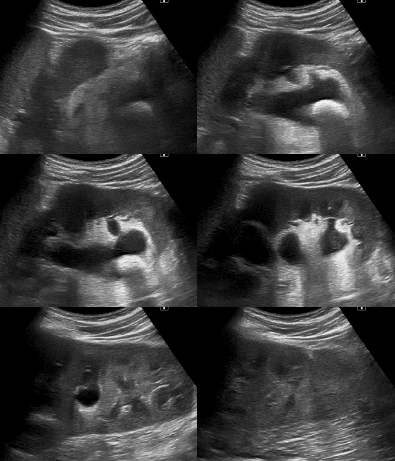
Urinary outflow obstruction will cause the pyelocaliceal system to dilate: this is termed hydronephrosis (fig. 36).
Hydronephrosis may have several causes, e.g. a lodged kidney stone, UPJ stenosis (fig. 37), or a lump exerting a mass effect on the ureter, or bladder retention (in reflux)
Kidney stones
Like bile stones, kidney stones cause acoustic shadowing (fig. 38). Very small renal concrements of a few millimeters may be missed in ultrasound examination as the acoustic shadowing is invisible. The gold standard to demonstrate or exclude nephrolithiasis is an abdominal CT examination without contrast.
Renal lesions
One of the most common abnormalities in the kidneys are cysts (fluid-filled sacs). Cysts are generally asymptomatic. Uncomplicated cysts are thin-walled, have anechogenic content and posterior wall enhancement & increased sound transmission (fig. 39). For additional information on ultrasound artifacts, see the Ultrasound Technique course.
Cysts may vary markedly in size. It is important to carefully evaluate the cyst wall and exclude any solid components. In the event of solid components or multiple septations, additional characterization using abdominal CT is indicated (fig. 40).


No comments:
Post a Comment