Spiral scanning in MRI is unlike spiral scanning in CT where the x-ray tube is continuously rotating and data is continuously being acquired. In MRI the word "spiral" refers to the pattern of sampling k-space. In conventional imaging sequences including spin echo and gradient echo and in fast imaging sequences, a line or multiple lines of k-space in the frequency direction are acquired consecutively. In spiral scanning, k-space is acquired in a spiral trajectory. The entire k-space can be acquired during a single acquisition, or interleaved using more than one acquisition. This sequence allows faster image acquisition than the fast echo sequences but is slower than echo-planar imaging. Spiral scanning tends to have fewer artifacts than echo-planar imaging since adjacent points in k-space are acquired in close temporal proximity. The figures to the right show how the acquisition of data in k-space is done with conventional sequences and with spiral scanning.
Friday, 29 March 2019
PD weighted spin-echo images
Proton-density weight images are related to the number of nuclei in the area being imaged (number of hydrogen protons), as opposed to the magnetic characteristics of the hydrogen nuclei. They are produced from the first echo. PD weight images result when the contribution of both T1 and T2 contrast is minimized. They have a long TR (2000+ms) to minimize T1 differences because all tissues exhibit full longitudinal relaxation prior to the next 90 degrees RF pulse. They have a short TE (TE1, 20ms) to minimize T2 differences. High PD tissues appear bright.
Fast spin echo
Fast or turbo spin echo (FSE/TSE) is an adaptation of conventional spin-echo (SE) acquisition technique designed to reduce imaging time. It has largely supplanted the original spin-echo technique due to vastly improved imaging speed.
T2 weighted
T2 weighted image (T2WI) is one of the basic pulse sequences in MRI. The sequence weighting highlights differences in the T2 relaxation time of tissues.
Thursday, 28 March 2019
MRI sequences
An MRI sequence is a number of radiofrequency pulses and gradients that result in a set of images with a particular appearance. This article presents a simplified approach to recognizing common MRI sequences, but does not concern itself with the particulars of each sequence.
Thursday, 21 March 2019
What if the patient is unable to sign consent what do we do?
| What if the patient is unable to sign consent what do we do? |
he patient has sickle cell disease. Can they get IV contrast?
| he patient has sickle cell disease. Can they get IV contrast? |
The patient has multiple myeloma. Can they get IV contrast material?
| The patient has multiple myeloma. Can they get IV contrast material? |
If a patient has thyroid disease is iodinated contrast contraindicated?
| If a patient has thyroid disease is iodinated contrast contraindicated? |
If a patient has thyroid cancer is iodinated contrast contraindicated?
| If a patient has thyroid cancer is iodinated contrast contraindicated? |
. Who should get baseline serum creatinine levels before CT?
| Who should get baseline serum creatinine levels before CT? |
If a CT is done on a pregnant patient and iodinated contrast is used can that affect the fetus?
| If a CT is done on a pregnant patient and iodinated contrast is used can that affect the fetus? |
What is our policy for scanning a pregnant patient?
| What is our policy for scanning a pregnant patient? |
If a patient is nursing can she receive IV contrast?
| If a patient is nursing can she receive IV contrast? |
Can you use iodinated contrast on a patient with suspected or known pheochromocytoma?
| Can you use iodinated contrast on a patient with suspected or known pheochromocytoma? |
Is there any problem with using iodinated contrast for CT if a patient is on the cardiac drug Amiodarone?
| Is there any problem with using iodinated contrast for CT if a patient is on the cardiac drug Amiodarone? |
What are the current guidelines for patients taking metformin and needing a contrast study?
What are the current guidelines for Metformin and Iodinated Contrast agents?
|
If a patient has myathenia gravis should iodinated contrast be used for a chest CT?
| If a patient has myathenia gravis should iodinated contrast be used for a chest CT? |
If a patient is on glucophage (or other oral diabetes medications) is there an issue with iodinated contrast agents? What is the current rule with oral diabetes drugs? (glucophage)
| If a patient is on glucophage (or other oral diabetes medications) is there an issue with iodinated contrast agents? What is the current rule with oral diabetes drugs? (glucophage). |
What patients take glucophage (metformin)?
| Metformin is approved by the FDA for first line treatment of Maturity Onset Diabetes Mellitus (NIDDM) and is used after diet alone fails. |
What are the other names for metformin?
| These brand names include; Glucophage, Glucophage XR, Glumetza, Fortamet, Riomet, Glucovance, Metaglip, Avandamet and a few others. |
What is the danger of Metformin and iodinated contrast?
| Biguanides are known to cause lactic acidosis in patients with a predisposing risk factor. The actual incidence for metformin is, however, quite low and when it has occurred has frequently been in patients with risk factors. |
Are there specific patients or risk factors that make it more likely to get a contrast reaction?
| Predisposing risk factors for general acute adverse reactions to contrast media are: |
Will faster injection rates (5 cc/sec vs. 1 cc/sec) result in an increased incidence of contrast reactions?
"The prevalence of anaphylactoid reactions is not affected by the rate of injection." Ionic Versus Nonionic Contrast Media: A Prospective Study of the Effect of Rapid bolus Injection on Nausea and Anaphylactoid Reactions. |
What are the categories of contrast reactions?
| What are the categories of contrast reactions? | |
Three Categories:
Mild Reactions Include
Moderate reactions include
Severe reactions include
|
Who can not get IV contrast material?
| The list will vary from site to site but here are some rules of note. Please remember that this list is not complete but a reasonable guide for the user. |
What if extravasation does occur? How often are there severe complications?
| The good news is that with most contrast extravasations prompt treatment prevents most complications. The literature recently has a few publications that address this issue. |
do we treat contrast extravasation when it happens?
| Part 1: How do we treat contrast extravasation when it happens? Part 2: Is cold compresses the rule or hot compresses? |
How do you prevent contrast extravasation?
| The key is to anticipate what can go wrong and make sure it doesn’t. it’s the classic risk prevention strategy. |
What is contrast extravasation and how often does it occur?
Contrast extravasation occurs when contrast is injected into a vein but goes outside of the vein into the adjacent soft tissues. In most cases this is a mild inconvenience to the patient especially when only a few cc are involved. With larger extravasations (75-100 cc) problems can occur.
| |||||||||||||
The patient is allergic to shellfish or crabs. Can they get a IV contrast?
| The reaction to shellfish and crabs is different than the issues of IV contrast. Therefore if your allergy is solely to crabs, shrimp, or lobster you don’t need to worry about getting IV contrast.
For the rest of my recommendation-just don’t eat shellfish (perhaps G-d was right)
|
Our patient needs the study now. What else can we do?
| Intravenous hydrocortisone at 200 mg can be used but the ACR recommends at least a 6 hours before doing the study. |
premedicate these patients are allergic to IV contrast
Part 1: What patients are allergic to IV contrast?
Part 2: Can we premedicate these patients and if yes what is our premedication protocol?
|
What if a patient is allergic to IV contrast material?
Since non-ionic contrast has been widely utilized, the incidence of allergic reactions has decreased. However, some patients may still be allergic to IV contrast agents.
|
s it possible for a patient to get a delayed reaction to IV contrast?
| Yes. Delayed reactions occur from more than 30-60 minutes post injection to 1 week post injection. The majority occur between 6 and 12 hours. |
Allergic Reactions
1. Part 1: Can a patient get a rash from IV contrast? Can it occur 24-36 hours post CT study?
Part 2: How do you treat the rash?
|
What contrast do you put in the bladder (type and volume)?
| We drip in up to 530 cc of contrast (500 cc bag of saline mixed with 30cc of Omnipaque-350).
The contrast is dripped in under gravity and when the patient has pain or the contrast runs out we will stop and then start the study.
|
Do we ever place contrast in the bladder similar to a cystogram and if so why?
| In cases of suspected bladder perforation, be it due to trauma (MVA and commonly associated with pelvic fractures) or surgical complication, the most accurate technique to detect contrast extravasation is a CT Cystogram. The presence of perforation, its site and its cause can all be defined on this study. The key to a successful exam is distension by contrast of the bladder under gravity (contrast dripped in) and typically not by hand injection of the contrast. Bladder rupture can be classified as intra and extraperitoneal. The intraperitoneal cases typically require surgery. |
What is virtual colonoscopy and what is the patient prep for the study?
Virtual colonoscopy is a new exam that is used for screening the colon for polyps. It is usually done in asymptomatic patients as part of the American Cancer Society's screening program but is also used in patients with failed classic fiberoptic colonoscopy.
|
Do we ever use rectal contrast and if so when and why?
| We do use rectal contrast for select applications. The basic principle would be for cases where opacification of the rectum and sigmoid colon is needed for defining the presence and/or absence of specific pathology. Some of the specific applications include: |
Are there any contrast volume limitations for the use of IV contrast?
Indeed there is and here are some rules for Omnipaque and Visipaque courtesy of Rich Vitti MD from GE Healthcare.
|
Have you ever seen a patient develop diffuse erythema distal to the IV injection site in the absence of extravasation?
| Here is the answer courtesy of Richard A. Vitti MD from Medical Affairs at GE Healthcare.
Rare, isolated cases of localized redness at the injection site have been reported to us at GE Healthcare over the years since the introduction of our non-ionic Omnipaque and Visipaque products.
|
Is it ok for patients to have both an MR and a CT with contrast on the same day?
| This question was addressed on the RSNA site and there is no issue doing both MRI and CT the same day with Gadolinium and Iodinated Contrast. |
Is there a relationship between patients receiving chemotherapy and CIN?
The list a number of factors that increase the risk for CIN and chemotherapy is one of them.
|
. What are some of the common normal “side effects” of IV contrast agents?
By RSNA and the ACR notes that “When an iodine-based contrast material is injected into your bloodstream, you may have a warm, flushed sensation and a metallic taste in your mouth that lasts for a few minutes.”
Monday, 18 March 2019
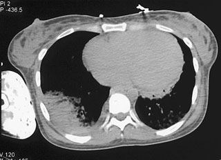
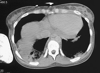
CT abdomen general
CT abdomen general
Indication/Technique
Please see the X-ray/CT Technique class for additional information about the technique of ‘computer tomography’ (CT).
Abdominal CT scans are generally evaluated in the transversal direction; the patient is seen from the feet upward as it were. The abdomen can also be viewed in the coronal and sagittal directions (fig. 1).
Abdominal CT scans are generally evaluated in the transversal direction; the patient is seen from the feet upward as it were. The abdomen can also be viewed in the coronal and sagittal directions (fig. 1).
Ultrasound Aorta
Aorta
Ultrasound is an effective and convenient way to confirm or exclude an abdominal aortic aneurysm (fig. 45).
Dimensions of the abdominal aorta:
Dimensions of the abdominal aorta:
- Normal < 2.5 cm
- Dilatation > 2.5 cm
- Aneurysm > 3 cm
In the event of abdominal aortic aneurysm, a CTA examination may be performed for additional evaluation.
CT protocol
No- for some studies we do a single phase acquisition and in some two or more phases. The different phases can potentially give more information than a single phase as well as to optimize lesion detectability. The phase usually refers to the time we acquire the scan data relative to IV contrast injection.
Common phases of data acquisition
- Arterial phase- 25-30 sec after injection
- Venous phase- 55-60 sec after injection
- Delayed phase- 240-300 sec after injection
Renal pathology
Renal pathology
Hydronephrosis
Urinary outflow obstruction will cause the pyelocaliceal system to dilate: this is termed hydronephrosis (fig. 36).
Gallbladder/bile duct pathology
Gallbladder/bile duct pathology
Bile stones
When the gallbladder contains bile stones, they can be imaged effectively using ultrasound. Bile stones are echogenic and opaque, causing “acoustic shadowing” immediately behind the stone (fig. 30).
Pathology
Pathology
- Liver pathology (steatosis, liver lesions)
- Gallbladder/bile ducts (bile stones, cholecystitis, dilated bile ducts)
- Renal pathology (hydronephrosis, kidney stones, kidney lesions)
- Bladder pathology (clot, bladder tumor)
- Spleen (splenomegaly)
- Pancreas (pancreatic tumor)
- Aorta (aneurysm)
- Intestines (appendicitis, diverticulitis, intestinal wall thickening)
- Trauma
Ultrasound Intestines
Intestines Ultrasound
The (small) intestines can never be imaged in their entirety by ultrasound. However, ultrasound may be very helpful in common intestinal pathologies.
The intestinal wall appearance changes markedly from the small intestine (Kerkring folds) to the colon (haustrations). Intestinal gas is a limiting factor in reliable evaluation of the intestinal wall (fig. 19).
The intestinal wall appearance changes markedly from the small intestine (Kerkring folds) to the colon (haustrations). Intestinal gas is a limiting factor in reliable evaluation of the intestinal wall (fig. 19).
Ultrasound Bladder
Bladder Ultrasound
The bladder must be filled for adequate evaluation. A filled bladder has thin walls with hypoechogenic content (fig. 13). A filled bladder creates a perfect acoustic window for evaluation of the distal ureteral orifices in the bladder posterior wall.
Ultrasound abdomen general
Indication/Technique
This course provides general information about ultrasound examination of the abdomen, with details on how it can and cannot be used.
Primary indications:
- Abdominal pain (including appendicitis, bile stones, kidney stones), herniation, umbilical hernia, inguinal hernia.
- Ascites
- Abdominal aneurysm
- Abnormal blood test results (liver and renal impairment)
- Unexplained fever
- Trauma (internal bleeding screening)
- Oncology (liver metastases screening)
Hip X ray
Indication/Technique
Indication
The hip X-ray is used primarily to demonstrate/exclude a fracture. Hip X-rays are also frequently opted for as initial test in chronic hip symptoms, e.g. osteoarthritis.
Technique
The hip joint can be imaged under various angles. A standard hip X-ray examination generally includes an anteroposterior (PA) image and a lateral image. Ideally, the AP image shows both hip joints (which strictly speaking makes it a pelvis X-ray) to allow comparison with the other hip. The lateral direction may be opted for in axiolateral images or a frog leg lateral image. The various directions are explained in more detail below.
Radiation protection
Due to the (potentially) negative long-term effects of radiation, the question whether the examination is indicated should be considered critically. Key questions include:
Harmful effects of radiation
Two harmful effects are distinguished:
- Deterministic effects; the likelihood and severity of the effects are dose-dependent. From a certain threshold dose, the body is no longer able to repair the radiation-induced cellular damage; think of red skin after prolonged radioscopy. Under the threshold dose, no effect occurs; above the threshold dose the severity of the effect increases with the radiation dose.
These are effects in the short term after high radiation dose.
Effective dose & types of examinations
The mean dose for conventional X-rays varies from 0.001 - 10 mSv and in CT tests from 2 – 20 mSv. For intervention procedures (radioscopy), the effective dose varies from 5 – 70 mSv and in nuclear medical procedures from 0.3 – 20 mSv.
Radiation
Radiation
his course will briefly address the (ionizing) radiation used in X-ray & CT examinations.
Most people know that (ionizing) radiation may be harmful to the human body. But why is this exactly?
Important questions such as ‘how do we measure radiation load?’, ‘how dangerous is radiation?’ and ‘how high is the risk of developing cancer when undergoing a CT examination?’ will be answered in this course.
Thursday, 14 March 2019
Reflection/deflection/absorption/scatter
Reflection/deflection/absorption/scatter
When sound waves move on the boundary surface between two media with different densities, part of the beam is reflected to the transducer. This phenomenon is called reflection. The remainder of the beam continues on into the tissue, but under a different angle. This is called deflection. As sound waves penetrate the tissue, part of the energy is converted into heat. This energy loss is called absorption. Finally, part of the sound waves are lost in scatter. This takes place when sound waves move through inhomogeneous tissue or in a 'hard’ boundary surface (= large density difference between two media). Part of the sound waves are reflected in random directions, a small part of which towards the transducer. For a summary see figure 10.
Ultrasound
Ultrasound Technique
General
Technique
- Transducers
- Frequency
- Different planes
- Reflection/deflection/absorption/scatter
- Color Doppler
- Duplex DopplerArtifacts
MRI contrast
MRI contrast
Frequent indications for MRI tests with contrast:
- Detect lesions (tumor/metastasis, abscess)
- Characterization of lesions (e.g. hepatic lesions)
- Imaging of vessels/vascular pathology (= MR angiography)
General MRI Terms
General MRI Terms
Signal intensity:
In MRI the terms low, intermediate and high signal intensity are used. Depending on the scan protocol, tissue is imaged as white (= high signal intensity), as a gray tone (= intermediate signal intensity) or as dark gray/black (= low signal intensity).
In MRI the terms low, intermediate and high signal intensity are used. Depending on the scan protocol, tissue is imaged as white (= high signal intensity), as a gray tone (= intermediate signal intensity) or as dark gray/black (= low signal intensity).
Relaxation
Relaxation
When the radiofrequent pulse is switched off, the protons will return to their original resting phase; the XY axis reverts back to the Z axis. This is termed relaxation. Two separate processes take place during relaxation: longitudinal relaxation (T1 relaxation) and transversal relaxation (= T2 relaxation). Again, these two processes are independent and should be regarded as two separate processes.
Monday, 11 March 2019


GFR and what it really means?
Estimating Glomerular Filtration Rate (GFR) and Creatinine Clearance (CrCl) in Health and Disease: Using Popular Formulae (September 2009)
What about the new “purple PICC/central lines” I hear about?
CR Bard has developed a PICC line designed for use for CT scanning with power injection up to 5 cc/sec.
Can we use a central line or a PICC line for injection?
Hospital policies will vary but at Hopkins we have several well defined policies.
Can any IV the patient has in place be used to inject the contrast material?
| A 18-20 g IV line can be used with the caveat that the line functions well on test injections and the line can be monitored during the injection of the contrast. |
Has there been any new developments in technology that may help us high injection rates in patients who can not tolerate an 18g needle (or at times even a 20g)?
The answer is yes. This past year the Nexiva Diffusics catheter got FDA approval for use.
What kind of IV access is ideal for use for IV contrast injection?
The best site for IV access is the right antecubital fossa.
What are the common volumes of contrast used for IV injection?
With the newest 64 MDCT scanners most patients have contrast volumes in the 100-120 ml range.
Should patients be NPO for CT scanning? If yes for how long?
We like the patient to be NPO for food for a minimum of 3 hours. However, fluids are encouraged (water is best) to keep the patient well hydrated. Dehydration is a leading factor for development of CIN.
. Can we pretreat patients who have borderline renal function? If yes then how?
Two common regimens are the use of bicarbonate in solution as part of a prestudy ( and often post study protocol) hydration protocol.
Do we have set cutoffs for creatinine levels and if so what are they?
| Although this will vary on the patient |
What patients are considered high risk patients for IV contrast for CIN?
This answer is very broad but here are some typical responses :
When do you use Visipaque-320 and when Omnipaque-350?
Our standard agent is Omnipaque-350 but for high risk patients we use an iso-osmolar agent which is Visipaque-320.
What is the advantage of Visipaque as written in the literature?
Summary from the American College of Cardiology publication 2012 ACCF/AHA/ACP/AATS/PCNA/SCAI/STS Guideline for the Diagnosis and Management of Patients With Stable Ischemic Heart Disease.
Why do you warm IV contrast?
With extrinsic warming of contrast there is a 3x less frequency of extravasation than if not warmed when high injection rates are used.
Are all CT scans with IV contrast done the same way?
| No- for some studies we do a single phase acquisition and in some two or more phases. |
3. What is GFR and why is it a more accurate measure than simply getting a creatinine level?
Use of GFR or glomerular filtration rate is becoming more common as a standard for evaluating patient prior to contrast media injection. |
Sunday, 10 March 2019
Use GFR practice
Do we use serum creatinine levels or GFR in practice for establishing risk prior to CT Scanning ?
Saturday, 9 March 2019
Time relate between Oral and actual scan
when does the patient get the oral contrast relate to the time patient get actual CT scan?
CT Upper Adbomen


CT upper abdomen with contrast Americans man with body weight 85kg high 1.75cm :
Contrast media : 100 ml Ultravist 300ml/mg
Follow rate : 2.5cc/ml
Technique. : 35 sec of arterial venouse 75sec
Advantage of Oral Contrast
What are the advantages of Oral contrast ?
1. Poorly absorbed from the normal GI tract.
2. Rapidly absorbed from peritoneum.
3.Rapidly dissipate from lung if aspirations.
4. Well accepted by patient due to neutral tastes .
1. Poorly absorbed from the normal GI tract.
2. Rapidly absorbed from peritoneum.
3.Rapidly dissipate from lung if aspirations.
4. Well accepted by patient due to neutral tastes .
Friday, 8 March 2019
Monday, 4 March 2019

Subscribe to:
Comments (Atom)