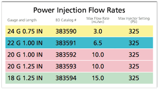
The answer is yes. This past year the Nexiva Diffusics catheter got FDA approval for use. This allow us to use smaller needles and still get high injection rates. The 20g Diffusics can inject up to 10 cc/sec and the 22g up to 6.5 cc/sec. This is a major step forward.
IV Catheter Safety
- Most IV catheters are pressure rated and usually the safety on the injector is for a pressure of 300 psi or less. Exceeding this can result in product leakage and or damage to the catheter and potentially injury to the patient
- Some of the newer generation IV catheters like the BD Nexiva Diffusics can be used to a psi of 325
- Please be certain that you are aware of what catheter you use and in practice it is ideal to use one brand only to prevent errors in setting the psi values in routine practice
"The purpose of this study was to compare the performance of a 20-gauge fenestrated catheter with an 18-gauge nonfenestrated catheter for i.v. contrast infusion during MDCT."
I.v. contrast administration with dual source 128-MDCT: a randomized controlled study comparing 18-gauge nonfenestrated and 20-gauge fenestrated catheters for catheter placement success, infusion rate, image quality, and complications.
Johnson PT, Christensen GM, Fishman EK.
AJR Am J Roentgenol. 2014 Jun;202(6):1166-70.
"A 20-gauge fenestrated catheter performs similarly to an 18-gauge nonfenestrated catheter with respect to i.v. contrast infusion rates and aortic enhancement levels and can be placed in most subjects whose veins are deemed insufficient for an 18-gauge catheter."
I.v. contrast administration with dual source 128-MDCT: a randomized controlled study comparing 18-gauge nonfenestrated and 20-gauge fenestrated catheters for catheter placement success, infusion rate, image quality, and complications.
Johnson PT, Christensen GM, Fishman EK.
AJR Am J Roentgenol. 2014 Jun;202(6):1166-70.
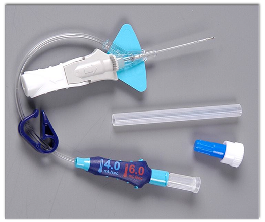
Hardware: IV Access Device
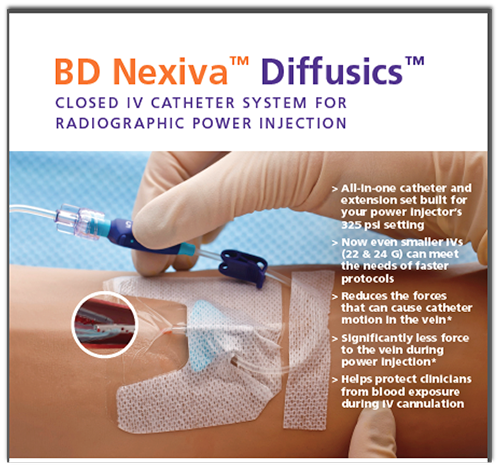
BD Nexiva Diffusics
Injection Rates
BD Nexiva Diffusics
"Experienced IV starters usually achieve IV access in one attempt by tailoring IV catheter gauge to vein quality; however, target infusion rates are not likely to be achieved with 22- and 24-gauge catheters, used in nearly 1/3 of the patients in this study."
Catheter insertion for intravenous (IV) contrast infusion in multidetector-row computed tomography (MDCT): defining how catheter caliber selection affects procedure of catheter insertion, IV contrast infusion rate, complication rate, and MDCT image quality.
Johnson PT, Christensen G, Lai H, Eng J, Fishman EK.
J Comput Assist Tomogr. 2014 Mar-Apr;38(2):281-4
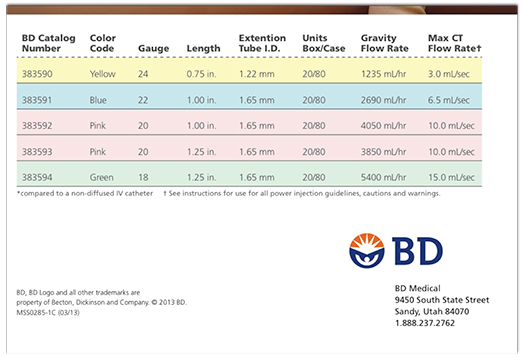
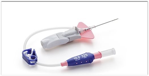
20 G Nexiva Diffusics
10.0 cc/sec injection rate
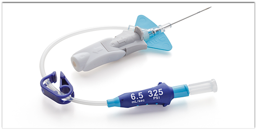
22 G Nexiva Diffusics
6.5 cc/sec injection rate
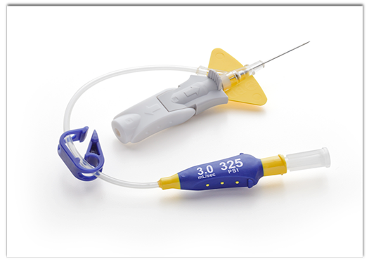
24 G Nexiva Diffusics
3.0 cc/sec injection rate
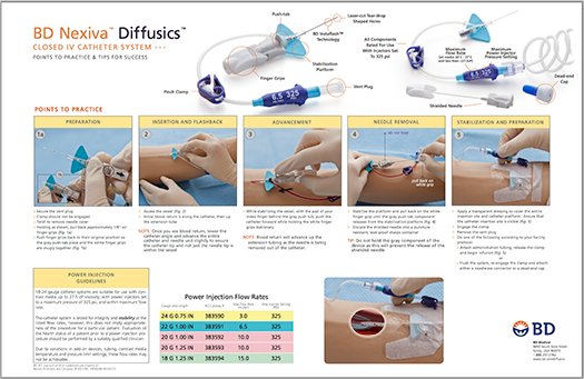
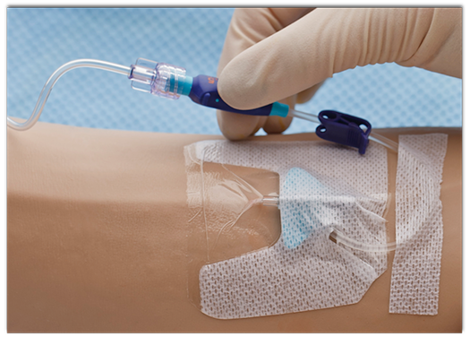
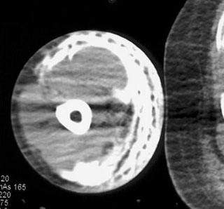
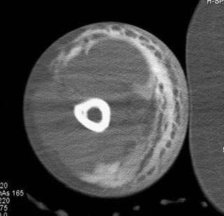
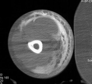
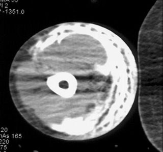
Nexiva in Use
Standard 18g vs Nexiva 20g
Nexiva Kit